Tuesday, 20 April 2010

Gamma amino butyric acid (GABA) and its receptor

GABA
GABA is the major inhibitory amino acid transmitter of the mammalian central nervous system and it is present in some 40% of all neurones.
Synthesis, storage and function
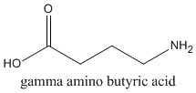
Gamma amino butyric acid (GABA) is the primary inhibitory neurotransmitter in the central nervous system. It is found in almost every region of brain, and is formed through the activity of the enzyme glutamic acid decarboxylase (GAD).

GAD catalyzes the formation of GABA from glutamic acid. The synthesis of GABA is linked to the Kreb's cycle. GAD requires vitamin B6 (pyridoxal phosphate) as a cofactor, which can be used to regulate the levels of GABA. GABA is destroyed by a transamination reaction, in which the amino group is transferred to alpha-oxoglutaric acid (to yield glutamate), with the production of succinic semialdehyde, and then succinic acid. The reaction is catalysed by GABA transaminase.
Vigabatrine, a GABA agonist, is used to treat epilepsy by inhibiting GABA transaminase.
GABA-ergic neurons and astrocytes take up GABA by specific transporters, and it is this, rather than GABA transaminase, that removes the GABA after it has been released. GABA transport is inhibited by Guvacine and Nipecotic acid.
Compounds such as the competitive GAD inhibitor allylglycine, inhibit GABA formation and cause convulsions due to the lack of GABA activity.
Sodium valproate (or valproic acid) on the other hand, blocks GABA transaminase activity, thereby elevating GABA levels, and thus alleviating seizures. Sodium valproate is useful in the treatment of epilepsy and bipolar mood disorders. Another strategy is to block GABA-transaminase with g-acetylenic GABA, thereby increasing the concentration of GABA at the synapse.

GABA receptors

Structure and Pharmacology
The GABA receptors are a class of receptors that respond to the neurotransmitter gamma aminobutyric acid (GABA), the chief inhibitory neurotransmitter in the vertebrate central nervous system. There are three classes of GABA receptors: GABAA, GABAB, and GABAС.
GABAA and GABAС receptors are ligand-gated ion channels (also known as ionotropic receptors), whereas GABAB receptors are G protein-coupled receptors (also known as metabotropic receptors).

Ligand-gated ion channels

GABAA
It has long been recognized that the fast response of neurons to GABA that is blocked by bicuculline and picrotoxin is due to direct activation of an anion channel.
This channel was subsequently termed the GABAA receptor. Fast-responding GABA receptors are members of family of Cys-loop ligand-gated ion channels. Members of this superfamily, which includes nicotinic acetylcholine receptors, GABAA and GABAС receptors, glycine and 5-HT3 receptors, possess a characteristic loop formed by a disulphide bond between two cysteine residues.
The GABAA receptor consists of three separate subunits with an alpha2, beta2, gamma arrangement similar to that found for neuronal nicotinic receptors. The ligand binding site is located at the interface between the alpha and beta subunits. The benzodiazepine binding site is located at a similar level at the interface between the alpha and gamma subunits. Each subunit is comprised of four hydrophobic sequences which span the membrane, with a large extracellular amino terminus containing the binding site and a carboxyl terminus located intracellularly.
Electrophysiological studies demonstrated that the activation of the receptor resulted in increased chloride conductance of the cell membrane with the concentration-response curve exhibiting positive cooperativity, consistent with the presence of at least two agonist binding sites on the receptor molecule. The agonist induced current decreased on continued exposure to high agonist concentrations suggesting that these receptors undergo desensitisation.

GABAC
In addition to the GABAA receptors there is a distinct class of ligand gated ion channels that are activated by GABA; referred to as the GABAC receptor. The natural agonist GABA is about an order of magnitude more potent at the GABA receptors than at the most common of the GABA receptors. The GABA receptors are activated by cis-aminocrotonic acid (CACA), which is not recognised by either the GABAA or GABAC receptors, suggesting that they recognise the partially folded conformation of GABA.
GABAC receptors are not blocked by bicuculline and do not recognise the benzodiazepines, barbiturates or the neuroactive steroids but, like GABAA receptors are blocked by picrotoxin, while 1,2,5,6-tetrahydropyridine-4-yl methyl phosphinic acid appears to inhibit GABAC receptors selectively.
However, molecular cloning studies have revealed that this pharmacological profile is remarkably similar to that exhibited by the rho-subunits when expressed ectopically. Two homologous subunits, rho1 and rho2, have been identified in man and these can be expressed as homomers or heteromers, but do not co-assemble with any of the GABA receptor subunits.

Common characteristics
In ionotropic GABAA and GABAС receptors, binding of GABA molecules to their binding sites in the extracellular part of receptor triggers opening of a chloride ion-selective pore.
Opening of a chloride conductance drives the membrane potential towards the reversal potential of the Cl¯ ion which is about –80 mV in neurons, inhibiting the firing of new action potentials.
However, there are numerous reports on GABAA receptors, which are actually excitatory. This phenomenon is due to increased intracellular concentration of Cl¯ ions either during development of the nervous system or in certain cell populations.
After this period of development, a Chloride pump is up-regulated and inserted into the cell membrane, pumping Cl- ions into the extracellular space of the cell. Further openings via GABA binding to the receptor then produce inhibitory responses. Over-excitation of this receptor induces receptor remodeling and the eventual invagination of the GABA receptor. As a result, further GABA binding becomes inhibited and IPSPs are no longer relevant.

G protein coupled receptor: GABAB
A slow response to GABA is mediated by GABAB receptors, originally defined on the basis of pharmacological properties.
In studies focused on the control of neurotransmitter release, it was noted that a GABA receptor was responsible for modulating evoked release in a variety of isolated tissue preparations. This ability of GABA to inhibit neurotransmitter release from these preparations was not blocked by bicuculline, was not mimicked by isoguvacine, and was not dependent on Cl¯, all of which are characteristic of the GABAA receptor. The most striking discovery was the finding that baclofen (β-parachlorophenyl GABA), a clinically employed spasmolytic mimicked, in a stereoselective manner, the effect of GABA.
Later ligand-binding studies provided direct evidence of binding sites for baclofen on central neuronal membranes. cDNA cloning confirmed that the GABAB receptor belongs to the family of G-protein coupled receptors. Additional information on GABAB receptors has been reviewed elsewhere.

Drugs acting on GABA receptors
Below is the table showing the effector pathway, agonists and antagonists of GABA receptors.

GABAA
GABA binds to GABAA receptors in the extended form as demonstrated by the activity of trans 4-aminocrotonoic acid and the lower activity of cis 4-aminocrotonoic acid.
Muscimol is a naturally occurring compound isolated from Amanita muscaria and acts as a GABAA agonist. The isoxazole ring also is found in the GABAA agonist tetrahydroisoxazolopyridinol (THIP).
Bicucullin acts as a direct antagonist at the GABAA receptor as does SR 42641.

GABAB

Baclofen is direct agonist at GABAB receptors, which are coupled to G proteins. GABAB receptors may regulate Ca2+ and K+ influx through the Gi/o family of G proteins and act presynaptically to inhibit the release of excitatory amino acids such as glutamate. Baclofen is orally active as a muscle relaxant and has been used in the treatment of rigidity and spasticity of cerebral palsy. Phaclofen is a weak partial agonist, while saclofen is an antagonist at GABAB receptors.

Anxiolytics: Benzodiazepines
Benzodiazepines represent a class of compounds collectively referred to as anxiolytics. Benzodiazepines modulate the binding of GABA to the GABAA receptor. Benzodiazepines increase the binding of GABA to GABAA receptors and promote Cl- influx.
All the overt effects of the benzodiazepines: sedative, anxiolytic, anticonvulsant, muscle relaxant and amnesic, are produced via the GABAA receptors.
Diazepam (Valium) is one of the most widely prescribed sedatives on the market.

Benzodiazepines exhibit anxiolytic activity due to their ability to promote GABA binding. They act as indirect agonists. Several ligands can block the actions of benzodiazepines, including flumazenil, a benzodiazepine antagonist.

Anticonvulsant drugs
Anticonvulsants are used to treat the various forms of epilepsy, which is characterized by excessive neuronal firing in the cortical and temporal lobe regions of the brain. Electroencephalograms (EEG) can pick up the rhythmic discharge of neurons from electrodes placed on the scalp. Rhythmic discharges of spikes and slow waves characterize the EEG during seizures.
The benzodiazepines have an anticonvulsant action in addition to the anxiolytic activity. Recent work has helped distinguish between the two roles for the benzodiazepines. Picrotoxin is a convulsant which interacts with the GABA receptor complex and blocks the Cl- ionophore.
Diphenylhydantoin (phenytoin) is useful in the treatment of epilepsy. Phenytoin stabilizes the neuron against the excitation associated with seizure activity, without producing sedation.


Friday, 2 April 2010

Beta-adrenoceptors

রাজশাহী বিশ্ববিদ্যালয়ের ফার্মেসী বিভাগের এম. ফার্ম সিলেবাস অনুযায়ী প্রণীতঃ
Syllabus- Molecular and cellular mechanisms of 1) Glutamate receptors, 2) GABA and its receptors, 3) Catecholamine receptors ( alpha- and beta-adrenoceptors, dopamine receptors), 4) Acetylcholine receptors (nicotinic and muscarinic receptors), 5) Opioid receptors.

β-Adrenoceptor Subtypes

The β -adrenoceptors were initially divided into β1 and β2-adrenoceptors defined in terms of agonist potencies, β1-adrenoceptors demonstrated equal affinity for adrenaline and nor- adrenaline while β2-adrenoceptors displayed a higher selectivity for nor-adrenaline than for adrenaline. The discovery of these receptor subtypes led to the development of selective agonists and antagonists for each subtype.
The story does not end there; further experimentation using β-antagonists exposed another receptor subtype which appeared to be insensitive to typical β-adrenoceptor antagonists this was classified as β3-adrenoceptor. More recent pharmacological evidence is now emerging in support of a further receptor subtype β4-adrenoceptor, although as yet there are no selective compounds for this particular subtype.

Characteristics of β-adrenoceptors
β1 adrenoceptors
All β1 adrenoceptors mediate responses to noradrenaline released from the sympathetic nerve terminals and to circulating adrenaline. β1-adrenoceptors were originally defined based on their rank order of potency of these endogenous agonists and by the synthetic agonist isoprenaline. Isoprenaline has shown to be the most potent of the three agonists with adrenaline and noradrenaline showing equal affinity for this particular receptor.
Subsequently, synthetic antagonists have been identified for β1 receptors these include the non-selective propranolol as well as the more selective compounds like metoprolol, practolol, atenolol and betaxolol. The selective β1-adrenoceptor agonists so far identified (denopamine, xamoterol and Ro 363) have limited utility as pharmacological tools because of low selectivity and/or efficacy.

β1-Adrenoceptor Location and Function
β1-adrenoceptors are largely postsynaptic and are located mainly in the heart but are also found in platelets, the salivary glands and the non-sphincter part of the gastrointestinal tract (GIT). They can however be found presynaptically. Activation causes an increase in the rate and contractile force of the heart, relaxation of the non-sphincter part of the GIT, aggregation of platelets and amylase secretion from the salivary glands. Presynaptically, their activation causes an increase in noradrenaline release.
The major ligands used for the autoradiographic localisation of β-adrenoceptor subtypes are [125I] cyanopindolol, [3H] dihydroalprenolol and [125I] pindolol. Of these [125I] cyanopindolol is the most commonly used because of its high affinity, ease of preparation, relatively short exposure time, and selectivity for β-adrenoceptors.
Catecholamines acting on β1 adrenoceptors mediate a number of tissue responses including increases in cardiac rate and force of contraction, stimulation of renin secretion, relaxation of coronary arteries and relaxation of gastrointestinal smooth muscle.

Transduction Mechanisms
All known adrenoceptors are coupled to their effector systems by guanine nucleotide binding proteins (G proteins). β1 adrenoceptors are positively coupled to the membrane bound enzyme adenylate cyclase via activation of Gs G-protein. Activation of adenylate cyclase produces alterations in cellular activity by increasing intracellular levels of cAMP. In the myocardial cell increases in cAMP cause more L-type voltage sensitive Ca2+ channels to open in response to depolarisation. Calcium then enters the cell by this route with each action potential and causes an immediate rise in the concentration of intracellular calcium.
Raised levels of intracellular calcium stimulate further calcium release from the sarcoplasmic reticulum (see diagram above). Activation of the contractile machinery is due partly to this influx of calcium but mainly to the secondary release from the sarcoplasmic reticulum.
Both the heart rate (chronotropic effect) and the force of contraction (inotropic effect) are increased, resulting in a markedly increased cardiac output and cardiac oxygen consumption. The cardiac efficiency is reduced. Catecholamines can also cause disturbance of the cardiac rhythm, cumulating in ventricular fibrillation.

β2 adrenoceptors
β2 adrenoceptors are mainly associated with causing relaxation of smooth muscle in a variety of tissues (see diagram below). The β2 subtype has a high affinity for the endogenous agonist adrenaline. Synthetic β2 agonists include terbutaline, salbutamol, salmeterol and zinterol, all of which have proved therapeutically useful in the treatment of asthma. Selective β2 antagonist have also been identified these include butoxamine and ICI118 551.
Diagram adapted from Pharmacology (4th edition) Rang Dale and Ritter

β2-Adrenoceptor Location and Function
β2-receptors are also mainly postsynaptic and are located on a number of tissues including blood vessels, bronchi, GIT, skeletal muscle, liver and mast cell. Activation results in vasodilatation, bronchodilation, relaxation of the GIT, glycogenolysis in the liver, tremor in skeletal muscle and inhibition of histamine release from mast cells.
The location of β2 adrenoceptors can be identified using the radiolabelled ligand [3H] dihydroalprenolol or [125I] iodopindolol and its analogues.
β2 adrenoceptors have been cloned and expressed in a variety of species. Cloning was first achieved by screening hamster genomic libraries with oligonucleotides complimentary to peptide fragments of purified hamster lung β2 adrenoceptors. This yielded a clone that when expressed had functional and radioligand-binding characteristics consistent with those of the β2 adrenoceptors. Furthermore there appears to be only minor species differences between these clones, with approximately 87 to 93% overall amino acid identity.
Catecholamines acting on β2 adrenoceptors mediate a number of tissue responses. Bronchial smooth muscle is strongly dilated by activation of β2 receptors. Uterine smooth muscle responds in a similar way and β2 agonist are frequently used to delay premature labour. Skeletal muscle is also affected by adrenaline acting on β2 receptors though the effect is far less dramatic than that on the heart. The twitch tension of fast contracting fibres (white muscle) is increased by adrenaline, particularly if the muscle is fatigued, whereas the twitch of slow (red) muscle is reduced.
In man adrenaline and other β2 agonists cause a marked tremor, this is demonstrated in the shakiness that accompanies fear and excitement or the excessive use of β2 agonists (treatment of asthma being an example of this). β2 agonists also cause long term changes in expression of the sarcoplasmic reticulum proteins that control contraction kinetics and thereby increase the rate and force of contraction of skeletal muscle. Clenbuterol is a β2 agonist, which acts in this way and has been used illegally by "sportsmen" to enhance performance.
Histamine release is also inhibited by catecholamines acting apparently at β2 receptors on mast cells and the release of glucose from the liver is also controlled by β2 receptors leading to glycogenolysis.

Transduction Mechanisms
β2 adrenoceptors are also positively coupled to the membrane bound enzyme adenylate cyclase via activation of Gs G-protein. β2 adrenoceptor activation causes relaxation of smooth muscle through the activation of adenylate cyclase. Stimulation of adenylate cyclase produces alterations in cellular activity by increasing levels of cAMP. The resulting increased levels of cAMP activate protein kinase A, which phosphorylates and inactivates myocin light chain kinase (MLCK), the contractile machinery of smooth muscle.

β3 adrenoceptors
The β3 adrenoceptor has a profile quite distinct from that of β1 and β2 adrenergic receptors. Rodents, humans and other mammals share many of the characteristic β3 properties, although observable species-species differences have been identified. The naturally occurring variant of the human β3 receptor was correlated with hereditary obesity in Pima Indians, in Japanese individuals and in Western obese patients. Weight gain was also observed in female mice whose β3 gene had been disrupted.
These examples provided a picture of the important role of the β3 receptor in the regulation of lipid metabolism and as a possible target for drugs to treat certain forms of obesity.

β3-Adrenoceptor Location and Function
β3-adreceptors are expressed predominately in adipose tissue. Activation is proposed to be involved with noradrenaline induced changes in energy metabolism via lipolysis and thermogenesis.
Early antagonist/agonist studies of β adrenergic receptors suggested the existence of atypical responses clearly different from effects mediated by the β1 and β2 adrenoceptors. The new agonist BR37344 stimulated lipolysis in rodent brown adipocytes even in the presence of the antagonist propranolol. This and other discoveries led to the notion that additional receptors existed in fat, in the gut, in the heart and possibly in skeletal muscle. Cloning, sequencing and expression of the β3 gene in various species has now served to confirm the existence of this receptor.
The β3 adrenoceptor is composed of a single 408 amino acid residue peptide chain that belongs to the family of G-protein coupled receptors. The G-protein receptors are characterised by seven hydrophobic stretches of about 22 to 28 residues forming seven transmembrane segments. The transmembrane (TM) regions are linked with three intracellular and three extracellular loops. The amino acid (N) terminal of these receptors is located extracellularly and is glycosylated. The carboxy (C) terminal is intracellular and the case of the β3 receptors does not have any phosphorylation sites.

Transduction Mechanisms
For all β-adrenoceptors transduction is via G-proteins coupled to the intracellular second messenger adenylate cyclase. All β-receptors are positively coupled to adenylate cyclase via activation of Gs G-protein. However, activation of the β2 and β3-adrenoceptors results in stimulation or stimulation and inhibition of adenylate cyclase. Activation of the β1 and β4 receptor results in an increase in the formation of cAMP and the subsequent stimulation of cAMP-dependent protein kinase.
Comparing β3 with β3 receptors of other species revealed a high degree of sequence homology approximately 80-90% between human, bovine, rodent and canine. The human, monkey and bovine β3 receptors are closer to each other than any of the rodents sequences, in particular in transmembrane segment one.

Molecular Biology of β-adrenoceptors
Extensive studies on the molecular features of the β-adrenoceptors have been carried out and the β-adrenoceptor is now among the best defined of the G-protein-linked superfamily of receptors.
The human β-adrenoceptors (β1, β2 and β3) are derived from at least three distinct genes and comprise of 477, 413 and 408 amino acids respectively. All three β-adrenoceptors show high amino acid sequence homology for each receptor between species. For example the β1 receptor of the mouse and rat are 90% homologous to the human β1. Similarly the mouse β2 receptor is 95% homologous to the human β2. However there are marked sequence differences between the subtypes. The β1 shows 48.9% homology with β2 and 50.7% homology with β3, whereas β2 show 45.5% homology with the β3.
The G-protein superfamilies are characterised by their seven hydrophobic transmembrane spanning domains. Three extracellular and three cytoplasmic (intracellular) loops, with a glycosylated extracellular amine (N) terminus and cytoplasmic carboxy (C) terminus link the transmembrane domains. The seven transmembrane domains arrange in the membrane to form a "pocket".
A great deal of the research done to determine the functional domains of the β-adrenoceptors has been carried out on the β2 receptor. Many of these studies have involved the preparation and expression of site directed mutations (deletion or substitution) of particular amino acids and also the synthesis of chimeric receptors.

β1
Three amino acids-Leu110, Thr117 and Val120 in TM2 of the β1-AR were identified as major determinants for β1-AR selective agonists. TM4 is responsible for β1-selective binding of noradrenaline.

β2
Important residues involved in ligand binding also include Asp79, 113, and 130. Asp79 appears to be essential for the binding of high affinity agonist but is not required for the binding of antagonists. Asp113 located in the third transmembrane domain is vital for binding of both agonist and antagonist in the β2 receptor. Substitution of this residue with a Glu results in a marked decrease in the ability of the receptor to couple to adenylate cyclase. Substitution of Asp130 increases the affinity of the receptor for the agonist but reduces the ability of the receptor to couple to the G-protein.

Other important amino acids associated with binding include Ser203, 204 & 207 in TM5; Tyr326 and Phe289, 290 alteration of these residues by substitution resulted in an increased affinity for agonist with no effect on antagonist binding. Ser204 forms hydrogen bonds with the meta hydroxy group of catecholamines; similarly Ser207 interacts with the para hydroxy group group of catecholamines.
Chimeric receptor studies revealed that transmembrane domains six and seven were important in determining the specificity of antagonist binding. Asn293 in TM6 interacts with the β-OH group of β-AR ligands and is responsible for stereoselectivity.
Residues of the third intracellular loop are involved in the coupling of the receptor to the Gs-protein, where the deletion of amino acids 222-229 and 239-272 caused complete loss of coupling to adenylate cyclase.

β3
Computer modelling has defined an image of the β3 ligand-binding site. At least four of the seven transmembrane domains are essential for ligand binding (see diagram below). The amino acids that are involved were identified by site-directed mutagenesis and photoaffinty labelling these are Asp117 in TM3, a residue found to be essential for binding all biogenic amines. Ser169 in TM4, is thought to form a hydrogen bonds with the hydroxyl of the ethanolamine side-chain. Ser209 and Ser212 in TM5, also located in many biogenic amine receptors, are thought to form hydrogen bonds with the hydroxyls of the catechol side chain. Also Phe309 in TM6, involved in hydrophobic interactions with the aromatic ring of catecholamines. Two of the three TM domains are involved in Gs activation, TM2, which contain Asp83, and TM7, which contains Tyr336.

Attempts to explain why several β2 antagonists behave as β3 agonist have not yet been successful. β3-adrenergic receptors appear to contain less bulky residues, and could therefore accommodate the larger β1/β2 antagonists. Mutagenesis has been used to substitute the small Gly residue with the larger Phe residue at position 53 in the β3 receptor; this change however was not enough to convert the β3 agonist to an antagonist.
The Gs interaction site on β3 is situated in the intracellular region, mainly the membrane proximal regions of the second and third (i2, i3) intracellular loops and the carboxy-terminal domains. Several reports propose that the β3 receptor may be coupled to more than one second messenger system. It has been shown that β3 receptors in rat adipocytes interact with both Gs and Gi proteins. The β3 adrenergic receptor in the septum of the human heart has also been reported to link with the Gi protein, resulting in a negative inotropic effect. This is in contrast to previous observations that always associated β3-adrenoceptors with positive inotropism in the heart.
All of the β-adrenoceptors have a glycosylated extracellular site on the N terminus; these sites are not associated with binding or signal transduction however it is thought that they are important for insertion of the receptor into the membrane. β adrenoceptors also contain a number of Cys residues (Cys106, 184, 190 & 191) which are thought to form disulphide bonds between the hydrophilic extracellular loops, which stabilise the receptor conformation.

β4-adrenergic receptor
β4-receptors are localised in cardiac tissue with activation causing an increase in heart rate and force.

Clinical Uses
Adrenergic drugs are used in the treatment of a wide range of medical conditions. Including the use of β2-receptor selective agonists in the treatment of asthma and other related bronchospastic conditions examples of these drugs include salbutamol and salmeterol. Beta-blocker drugs are commonly used in the treatment of angina pectoris, cardiac arrhythmia and for the long-term treatment of patients who survive myocardial infarction. β-receptor antagonists have also been used as anti-hypertensive for a number of years. Beta -blockers have also proven useful in the treatment of conditions such as migraine, anxiety disorders, hyperthyroidism, alcohol withdrawal and when applied topically are useful in the treatment of glaucoma and ocular hypertension.

Wednesday, 10 February 2010

Alpha1- and alpha2- adrenergic receptor

রাজশাহী বিশ্ববিদ্যালয়ের ফার্মেসী বিভাগের এম. ফার্ম সিলেবাস অনুযায়ী প্রণীতঃ

Syllabus- Molecular and cellular mechanisms of 1) Glutamate receptors, 2) GABA and its receptors, 3) Catecholamine receptors ( alpha- and beta-adrenoceptors, dopamine receptors), 4) Acetylcholine receptors (nicotinic and muscarinic receptors), 5) Opioid receptors.

Alpha- adrenoceptor Subtypes

With the aid of pharmacological and molecular biological techniques the alpha-adrenoceptor subtypes were determined. alpha-adrenoceptors exist on peripheral sympathetic nerve terminals and are divided into two subtypes alpha1, and alpha2. These subtypes were at first classified by their anatomical location; alpha1 is found mostly postsynaptically, whilst alpha2 although typically sited presynaptically, can also occur postsynaptically. These initial subtypes were further divided into alpha1a, alpha1b, and alpha1d; and alpha2a, alpha2b, alpha2c, and alpha2d. This knowledge has led to the development of selective agonists and antagonists for each subtype.

alpha1-adrenergic receptor
alpha1-adrenoceptors are of particular interest therapeutically because of their important role in the control of blood pressure. All alpha-adrenoceptors consist of single polypeptide chains with 7 membrane spanning domains, and are members of the G-protein coupled receptor superfamily.

Classification
There are three subtypes of the alpha1 receptor: alpha1A, alpha1B, and alpha1D. Uppercase letters are used to denote 'functionally defined' subtypes while lowercase is used to denote 'molecularly defined' subtypes. Most tissues express mixtures of the three subtypes, but the relative expression levels have been found to be different in different reports. These subtypes appear to coexist in different densities and ratios, and in most cases responses to alpha1-adrenoceptor selective agonists are probably due to activation of more than one subtype.

Alpha-Adrenoceptor Location and Function:
Alpha1- adrenoceptors are found throughout the body, they are found in the brain where their functional role is not yet clear, they also play critical roles elsewhere in controlling contraction and growth of smooth and cardiac muscle. alpha1-adrenoceptors are found in both the central and peripheral nervous system.
>In the Central Nervous System they are found mostly postsynaptically and have an excitatory function.
>Peripherally they are responsible for contraction and are situated on vascular and on non-vascular smooth muscle. Alpha1-adrenoceptors on vascular smooth muscle are located intrasynaptically and function in response to neurotransmitter release. For non-vascular smooth muscle they can be found on the liver, where they cause hepatic glycogenolysis and potassium release. On the heart they mediate a positive inotropic effect. Cause relaxation of GI smooth muscle and decrease salivary secretion.

Drug affinity and selectivity
The affinities and selectivities of drugs for alpha1- adrenoceptor subtypes have been determined primarily by competition for radioligand binding to heterologously expressed recombinant subtypes. Most antagonists show little or no selectivity between the three known alpha1-adrenoceptor subtypes. However, a variety of drugs, including prazosin which has selectivity for alpha1A, with varying degrees of selectivity have been found. Studies suggest that noradrenaline and adrenaline activate all three alpha1-adrenoceptor subtypes with similar potencies, and that synthetic agonists show significant selectivity between the subtypes.

Functional domains on alpha1
Investigative research into the function of the various domains and/or amino acid residues of the adrenoceptors has produced a number of findings. The aspartate in the third transmembrane domain and the two serines in the fifth transmembrane domain that are conserved in all catecholamine receptors probably interacts with the protonated amine and two hydroxyls of the catecholamines. Using selective alpha1A agonists (e.g. oxymetazoline) Hwa et al (1995) used site directed mutagenesis to identify critical residues in alpha1A- and alpha1B-adrenoceptors that are responsible for apparent differences in agonist binding potency. The results showed that conversion of alanine to valine in the fifth transmembrane domain and leucine to methionine in the sixth transmembrane domain of the alpha1B subtype increased this receptors affinity towards the selective alpha1A agonists until its affinity became similar to that of the alpha1A subtype. These two residues are therefore critical in subtype selective agonist binding, and may interact structurally within the receptor. Other studies suggest that the fifth transmembrane domain and a portion of the second extracellular loop are critically important in subtype selective antagonist binding. This kind of evidence suggests that alpha1-adrenoceptor antagonists may bind near the surface of the receptor, rather than deep within the transmembrane domains like the agonists.

Transduction Mechanisms
All alpha-adrenoceptors use G-proteins as their transduction mechanism. Differences occur in the type of G-protein the receptors are coupled to. alpha1-adrenoceptors are coupled through the Gp/Gq mechanism, whereas alpha2-adrenoceptors are coupled through Gi/Go. Gp/Gq activates phospholipase C that phosphorylates phosphatidyl inositol to produce inositol triphosphate, and diacylglycerol. These compounds act as second messengers and cause release of calcium from intracellular stores in the sarcoplasmic reticulum, and activation of calcium channels respectively. They produce their effects by the release of calcium.
Alpha-adrenoceptors are G-protein coupled receptors. The alpha1 class of adrenoceptors belong to the Gq/11 type of G-protein. An agonist acting at the alpha1-adrenoceptor binding site causes Gq/11 to activate phospholipase C dependent hydrolysis of phosphotidyl inositol 4,5, biphosphate. The conversion of this compound by phospholipase C results in the generation of 1) Inositol triphosphate, and 2) Diacyl glycerol (DAG). 1) Inositol triphosphate acts to release calcium from intracellular stores in the sarcoplasmic reticulum. 2) Diacyl glycerol synergises with calcium to activate protein kinase C which phosphorylates specific target proteins in the cell to change their function.

Alpha1 adrenoceptors have been implicated in other signalling pathways including; calcium influx, arachadonic acid release, and mitogenic activity. alpha1-adrenoceptors may couple directly to activation of calcium channels in certain cells. Activation of alpha1-adrenoceptors leads to potentiation of a calcium current in a protein kinase C dependent manner.
Phospholipase A2 is an enzyme responsible for the release of arachidonic acid from phospholipids. alpha1B and alpha1D adrenoceptors have been shown to couple to phospholipase A2 and cause the activation of this enzyme through a pertussis toxin - sensitive pathway in CHO cells.
Mitogenic activity refers to cell growth and the mechanisms underlying it. G-protein coupled receptors, including alpha1-adrenoceptors have been shown to have mitogenic activity through mitogen activated protein kinase pathways.

 Alpha2-adrenergic receptor


Classification
There are at least 3 different subtypes of the alpha2-adrenoceptor within a species: alpha2A-, alpha2B- and alpha2C-adrenoceptors. Alpha2-adrenoceptors are usually found presynaptically. Presynaptic alpha2 receptors inhibit the release of noradrenaline and thus serve as an important receptor in the negative feedback control of noradrenaline release. Postsynaptic alpha2 receptors are also found.

Alpha2-Adrenoceptor Location and Function
Alpha2-adrenoceptors: are found in both the central and peripheral nervous system. They are found both pre- and postsynaptically and serve to produce inhibitory functions.

-Presynaptic alpha2 receptors inhibit the release of noradrenaline and thus serve as an important receptor in the negative feedback control of noradrenaline release.
-Postsynaptic alpha2 receptors are located on liver cells, platelets, and the smooth muscle of blood vessels. Activation of these receptors causes platelet aggregation, and blood vessel constriction.

Sympathetic nerves are present at the adventitial-medial border of arteries and increase of noradrenaline at these sites causes constriction of the arteries. alpha2-adrenoceptor agonists as well as alpha1-adrenoceptor antagonists are therefore used for the treatment of hypertension. Blockade of presynaptic alpha2-adrenoceptors enhances the overflow of noradrenaline from sympathetic nerves and potentiates the response to sympathetic stimulation. This can be a problem when trying to functionally study innervated alpha2-adrenoceptors because alpha2-adrenoceptor antagonists, by inhibiting pre-junctional alpha2-receptors, also increase neurotransmitter release and thereby mask any contribution made by post-junctional alpha2-adrenoceptors.

Functional domains on alpha2
Alpha2-adrenoceptors are of comparable size to the beta-adrenoceptors but differ in structure from alpha1 and beta- by having relatively short amino and carboxyl termini, and by possessing a very long third intracellular loop. A few amino acid residues appear to be critical for agonist or antagonist binding. For example, if Phe412 of the alpha2A is mutated to asparagine, the affinity for several alpha2-adrenoceptor antagonists is reduced by several orders of magnitude. An aspartic acid in transmembrane helix 3 has been found to be neccessary for specific binding of ligands to alpha2-adrenoceptors. This was shown to by inducing a mutation in which Asp113 was substituted by asparagine, this resulted in the elimination of specific binding of [3H]yohimbine to the alpha2-adrenoceptor. Analysis with photoaffinity probes has shown that partial agonists and antagonist ligands bind to an amino acid within the fourth transmembrane-spanning helix, although the precise location of the attachment could not be determined.

Transduction Mechanisms
All alpha-adrenoceptors use G-proteins as their transduction mechanism. Differences occur in the type of G-protein the receptors are coupled to. alpha1-adrenoceptors are coupled through the Gp/Gq mechanism, whereas alpha2-adrenoceptors are coupled through Gi/Go. The alpha2-adrenoceptor G-protein, Gi/Go, has been shown to be negatively coupled to adenylate cyclase and so reduces the formation of cyclic AMP which leads to a decreased influx of calcium during the action potential - the ion responsible for transmitter release. Therefore lowered levels of calcium will correspondingly lead to a decrease in transmitter release. The alpha2-adrenoceptors belong to the Gi type of G-protein which acts to inhibit adenyl cyclase the enzyme responsible for synthesising the second messenger molecule cAMP from ATP. cAMP acts by activating protein kinases which catalyse the phosphorylation of serine and threonine residues in different cellular proteins, using ATP as the source of the phosphate groups. This mechanism acts to regulate cellular functions. The cellular functions cAMP can regulate include: cell division and cell differentiation, ion transport, ion channel function which leads to changes in electrical excitability, the contractile proteins in smooth muscle, and regulation of enzymes involved in energy metabolism.

The activation of an alpha2-adrenoceptor by agonist causes the alpha2-adrenoceptor to interact with a Gi type of G protein which inhibits the action of adenyl cyclase and thus the actions of cAMP.

Clinical Uses
The clinical uses of adrenergic compounds are vast. The treatment of many medical conditions can be attributed to the action of drugs acting on adrenergic receptors. alpha-adrenoceptor ligands can be used in the treatment of hypertension. Drugs such as indoramin and prazosin are alpha1-adrenoceptor antagonists and have antihypertensive effects, as is clonidine an alpha2 adrenoceptor agonist. alpha1-adrenoceptor antagonists are also employed in the control of benign prostatic hypertrophy. However there can be cardiovascular side effects associated with alpha1 block. Alpha2-adrenoceptor agonists such as clonidine are often used as an adjunct to general anaesthetics.